X-rays and scans take pictures of various parts of the body. Bones show up most clearly but you can also see other tissues.
Key points to remember
- x-rays and scans don't hurt but to do them properly your child will need to lie still
- an anaesthetist may give an anaesthetic or some sedation to help them stay still
What are x-rays?
X-rays take pictures of various parts of the body. Bones will show most clearly, but you can also see other tissues.
X-rays and scans don't hurt but to do them properly your child will need to lie still. This can be uncomfortable, especially if it is for a long time. An anaesthetist may give an anaesthetic or some sedation for this reason.
Bone scan
Staff at the nuclear medicine department run bone scans to get an image of a bone or bones. They study the image to see if your child has cancer in their bones. The scan itself is not painful.
A doctor or nurse will inject radioactive material (an isotope) before running the scan a couple of hours later. Areas that show too much tracer may mean cancer.
Computerised axial tomography (CT or CAT scan)
A CT scan uses a special x-ray to make a detailed picture of the inside of the body. Scanning happens in the radiology department.
A doctor or nurse may either:- inject contrast dye into your child's vein, or
- send you the contrast dye, with instructions, before the CT scan - you can give this to your child by mouth (orally)
Contrast dye helps recognise normal structures from cancer.
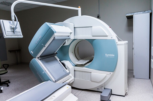
Magnetic resonance imaging (MRI)
An MRI uses a special machine (scanner) to look inside the body. The scanner uses magnetic waves to create a picture of the inside of the body.
Your child will need to lie still on the table inside the MRI machine during the scan. Your child should not wear anything metal (jewellery, belt, etc) because the machine attracts metal. While the MRI is scanning your child will hear and feel a rhythmic knocking sound, like a drumbeat.
You may not be able to stay in the same room with your child during this test but you and the staff can always hear and see your child.
The length of the test will depend on what part of your child’s body is scanned. Some children need sedation to lie still for the whole test.
MIBG scan (meta-iodobenzylguanidine scan)
MIBG scans help locate and diagnose certain types of tumours in the body. Neuroblastoma is one example of a tumour commonly identified using a MIBG scan.
A doctor or nurse will inject a small amount of radioactive tracer into your child's vein. After the injection, the scanner will take pictures. The scanner is similar to a CT scan.
The scans can happen 24, 48, and 72 hours after the tracer injection. A doctor or nurse will give your child a special medicine before and after the scan. This is to protect their thyroid gland from the radioactive substance in the tracer. Ask your healthcare team about how to prepare your child for the MIBG scan.
PET scan (positron emission tomography scan)
PET scans look for tumour activity in the body. They can also show infections or inflammation.
Before the scan, a doctor or nurse will inject a small amount of radioactive isotope, or tracer, into a vein. The tracer travels to places in the body where there is tumour activity. After the tracer injection, your child will have to lie very still on the PET scanner table while the scanner takes pictures.
Ask your healthcare team about what instructions your child needs to follow to prepare for the PET scan. There are special dietary instructions to follow, and there may be other instructions if your child needs sedation or a general anaesthetic.
Ultrasound
An ultrasound makes pictures of the inside of the body by bouncing sound waves off tissue or organs inside the body.
A doctor or radiographer will put a clear jelly on the relevant part of your child's body. They will then move a small round probe (transducer) around to get a clear picture of the tissue or organ.
Click Donate to fund the ongoing development of the Circulating Tumor DNA project which will improve outcomes for those children already diagnosed with cancer.
Disclaimer - Content on this blog is for educational use only. Please consult your doctor or other health professional to make sure the information is right for your child.
Copyright - Please note that the information on this blog is copyright owned by The Paediatric Society of New Zealand and Starship Foundation. See copyright information at the Kids Health website.